Development and design of medical endoscope
The application of endoscopes is quite extensive in the treatment of ENT (otolaryngology)-related diseases. The endoscope we designed for use in the ear can well meet the current otology applications, and the designed resolution class can reach 100lp/mm. The image is clear and can fully meet the medical application of ear science. The main function of the endoscope is to determine and observe the diseased tissue, and carry out early diagnosis and treatment [1-2]. Endoscopy is widely used in the diagnosis and treatment of ENT (otolaryngology) diseases [3-4]. Early ENT instruments include otoscopes, indirect nasopharyngoscopes, indirect laryngoscopes, etc., which belong to the first generation of products. The second-generation products are formed by integrating these equipment and equipment.
With the development of science and technology, a digital high-definition ENT endoscopy diagnosis and treatment workstation has appeared, which can provide high-definition ENT endoscopy video. In recent years, ear endoscopy has been more and more widely used in otological clinical medicine. It puts the observation probe into the ear, and under the illumination of its own light source, it has an imaging lens to capture the details of the ear, image it on a CMOS or CCD image sensor, and send it to the monitor after photoelectric conversion and image signal processing, showing clearly The enlarged image is for the doctor to observe. In the current market, most ear endoscopes are agents of foreign companies, and there are very few self-developed ear endoscopes.
1. Design ideas
1.1 Selection of initial structure
Only a reasonable initial structure selection can get a good lens, and it directly affects whether the design can be carried out smoothly. There are two methods for designers to choose. One is to design an initial structure using the principle of paraxial optics through the designer's experience, and then gradually adjust the structural parameters to obtain the required results. However, it is very difficult to rely on the designer to create the initial mechanism, and the designer needs to have certain work experience and rich theoretical reserves. Another method is to select a suitable initial structure for optical design in relevant literature and patents, and then optimize it. The initial structure of this design uses a US patent as a design starting point. The selection principle of the initial structure is that the aperture value is the same as the field of view and the design index requirements, and the focal length can be realized by scaling the lens size.
2. Design process
2.1 Input of the initial structure After selecting the corresponding initial structure, it is necessary to modify the various indicators of the initial structure. Through the zoom of the focal length and the input of the wavelength, field of view, and F number, the initial structure can meet a basic size requirement. . First zoom the focal length to 1.3mm, and then set the F number to 7.65. In this paper, the field of view is used to control the field of view, and the field of view and wavelength and the lens parameters of the initial structure are input in Zemax [5].
3. Design results
The optimized shape structure and system parameters of the lens are shown in Figure 1 and Table 1. The system is composed of 10 lenses, including two sets of doublet lenses, two double-convex lenses, one meniscus lens, one double-convex lens, and one flat lens. Among them, the glass materials from the first piece to the last piece are: H-ZF62, H-LAF10L, H-LAK53A, H-ZLAF75A, H-ZLAF53A, H-LAK2, H-ZF7LA, K9, F5, BAF8. The combination of crown glass and flint glass is good for correcting aberrations.
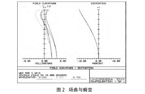
3.1 Field curvature and distortion
The field curvature reflects the curvature of the image plane of the entire optical system. For this type of endoscope lens, the field curvature should be less than 0.2 mm. It can be seen from Figure 2 that the field curvature correction is within 0.05 mm. In addition, for this low-cost endoscopic lens, the distortion requirements are relatively low. It can be seen from Figure 2 that the distortion of the peripheral field of view is within 10%, and at the 0.7 field of view, the distortion is about 5%, which meets the design requirements.
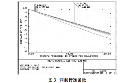
3.2 Modulation transfer function
The modulation transfer function (MTF) is the degree of attenuation of the contrast (that is, the amplitude) of the sinusoidal intensity distribution function of various frequencies after being imaged by the optical system. For the visual system, the threshold of the human eye is 0.3, and for the camera system, the threshold is 0.1. As shown in Figure 3, at 100lp/mm, all fields of view are greater than 0.3. Meet the design requirements [7-8].
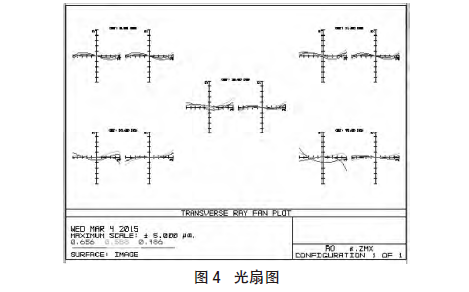
3.3 Optical fan diagram
The light fan diagram is about the ray aberration of the pupil coordinate function, and the data drawn is the difference between the coordinates of the intersection point of the ray and the image plane and the coordinates of the intersection point of the chief ray and the surface. They can well reflect the actual convergence of light on the image plane [9]. As shown in Figure 4, the light convergence is relatively good.
3.4 Spot diagrams
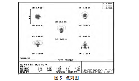
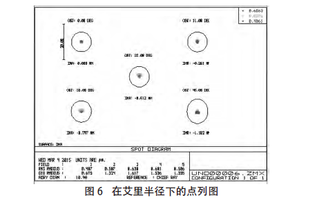
From Figure 5 and Figure 6, it can be seen that the root mean square radius of the imaging diffuse spot in each field of view of the system is much smaller than the radius of the Airy disk, and the energy is relatively concentrated, which meets the design requirements.
3.5 Relative illuminance
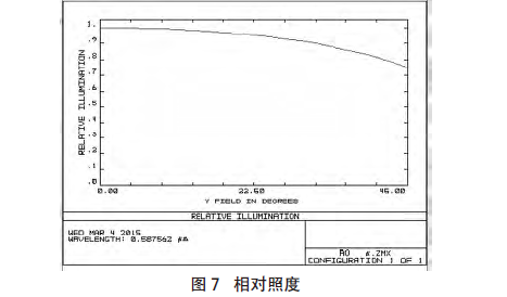
Relative illuminance refers to the ratio of the illuminance at the edge of the field of view to the illuminance at the center, and the higher the ratio, the brighter the edge. Generally speaking, relative illumination above 50% is acceptable. It can be seen from Figure 7 that the relative illumination of this lens is above 70%, which meets the design requirements.
4. Tolerance Analysis
4.1 Tolerance analysis method
In order to improve the imaging quality of this optical system, all parameters in the system need to be assigned variable tolerances. If the system fluctuates greatly or is more sensitive to changes in one of the parameters, the system needs a relatively high level of performance. Therefore, for The tolerance requirements of this group should be tight, whereas looser tolerances can be applied. Sight glasses have relatively high requirements for imaging, so the requirements for the tolerance of the optical system are relatively strict. Use the tolerance calculation and analysis program in the ZEMAX software to calculate the sensitivity of performance degradation of various parameters in the optical system, that is, to analyze the processing and assembly tolerances of all components.
4.2 Results of Tolerance Assignment
Using ZEMAX optical design software, through sensitivity analysis, inversion sensitivity analysis and Monte Carlo analysis, the reasonable tolerance distribution of the microscope objective lens is obtained. By calculating and analyzing the MTF drop of each tolerance parameter at the Nyquist spatial frequency of 100lp/mm, the appropriate tolerance is finally determined. The results of sensitivity tolerance analysis and Monte Carlo tolerance analysis are shown in Table 2 and Table 3 respectively. The Monte Carlo tolerance analysis results show that more than 90% of the Monte Carlo samples of the microscope objective system have MTF0.156524163, and each sample is a simulated processed and adjusted optical system.
Summary
By establishing the ideal initial state and initial structure of the optical system, the Zemax optical design software is used to optimize the structure design, and then an endoscopic lens that meets industrial standards and can be produced is obtained. Compared with other similar lenses, this lens has better distortion control at a 90° field of view, and a resolution of 100lp/mm, which is more conducive to observing diseased tissues. In summary, the endoscope meets medical needs.
Proposal recommendation
- TOP


